Monday, November 25, 2013

Day 40 - November 25, 2013

It has been 6 days since my last observation and quite a few things have changed within my Microaquarium. As mentioned in my last blog post, a second beta food pellet was added after my last observation 6 days ago and I can say with certainty that it has significantly affected the ecosystem in my Microaquarium. Today I was able to indentify, with Dr. McFarland's help, three new organisms that I hadn't identified in my previous observations, and in general, the abundance and diversity of organisms has increased significantly. This week for the first time I was able to indentify an additional cyclops of equal age but opposite gender to the one previously observed. I also observed a very juvenile cyclops. In addition to the newly identified organisms I was able to observe a swarm of bacteria in my Microaquarium. As I watched, huge numbers of various bacteria of different shapes moved very rapidly and in a sort of collective fashion forming a swarm. The swarm was limited to a vague cylindrical shape, and the whole collection of bacteria seemed to be moving to the right, in affect sweeping the area. Dr. McFarland postulated that it might be related to a biofilm.

Overall it has been amazing to see the ecosystem change over the last few weeks, and witness an environment and the evolution of a food chain first hand. It's been an incredible opportunity to use the equipment of the lab in Hesler biology and see these microscopic processes that take place largely without our knowledge. I have to thank Dr. McFarland for being so accommodating, and meeting with me outside of the normal observation hours.

Picture 1. Cyclops sp. A fully developed cyclops that is probably a male (Pennak, 1953, p. 384 Figure 241).


Picture 2. Raphidiophrys sp. A raphidiophrys, an amoeba, can be seen with microtubule structures extending from its center (Patterson p. 173 Figure 193).


Picture 3. Centropyxis sp. A centropyxis, an amoeba can be seen (Patterson p. 95 Figure 184).

In addition to the organisms pictured above, I was able to identify an Amoeba Radiosa (Patterson p. 80 Figure 142).  I was also able to observe an Epalxis sp. (Pennak p. 141 Figure 124).

The following video depicts the bacteria swarm:


Wednesday, November 20, 2013

Day 33 - November 18, 2013

It has been a few weeks since my last observation on Day 15, and a few interesting things have transpired during that time period. Principly, the cyclops has developed egg sacks and can now be assumed to be a female. Additionally, I observed some interesting phenomena; the anisonema that I saw previously has begun to attack worm castings, and the mosses have continued to deteriorate. Entire lengths of moss have now died and are covered in what appear to be fungal hyphae. This hyphae could not be indentified by Dr. McFarland or myself, but is never-the-less interesting and quite beautiful.

In addition to my observations this week, I added an additional beta food pellet.
On Monday November 18, 2013 "ONE" Beta Food Pellet was inserted into my microaquarium. "Atison's Betta Food" made by Ocean Nutrition, Aqua Pet Americas, 3528 West 500 South, Salt Lake City, UT 84104. Ingredients: Fish meal, wheat flower, soy meal, krill meal, minerals, vitamins and preservatives. Analysis: Crude Protein 36%; Crude fat 4.5%; Crude Fiber 3.5%; Moisture 8% and Ash 15% (McFarland 2013).

It will be interesting to see how the additional food pellet affects both the organisms living in the Microaquarium and the system itself. I suspect the first beta food pellet added to the environment contributed to the decline in quality and clarity of the water, and I wouldn't be surprised if the second causes a similar result.

This week I used the same videomicroscope I've used for my previous observations.


Picture 1. Cyclops sp. An older cyclops that has developed egg sacks and can now be identified as a female. (Pennak, 1953, p. 384 Figure 241).


Picture 2. An unidentified organism growing on a dead length of Fontinalis sp. (McFarland 2013). Assumed to be a species of fungal hyphae.


Saturday, November 2, 2013

Day 15 - October 31, 2013

Since my last observation on Day 9, my MicroAquarium hasn't had any significant change. Most notably, the mosses and carnivorous plant have detriorated in health significantly. Parts of the leaves and stems of the plants have died and the colors of all three have taken on a significantly brown hue. I can assume this is because of continued degradation of the water in my MicroAquarium, and maybe a declining oxygen concentration.

On Friday October 25, 2013 "ONE" Beta Food Pellet was inserted into my microaquarium. "Atison's Betta Food" made by Ocean Nutrition, Aqua Pet Americas, 3528 West 500 South, Salt Lake City, UT 84104. Ingredients: Fish meal, wheat flower, soy meal, krill meal, minerals, vitamins and preservatives. Analysis: Crude Protein 36%; Crude fat 4.5%; Crude Fiber 3.5%; Moisture 8% and Ash 15% (McFarland 2013).

It's also possible that the food pellet that was added to my MicroAquarium has contributed to the pollution of the system or at least increased the relative number of particulates in the water. This could be interfering with the mosses and carnivorous plants health.

I got to use the same videomicroscope this week as I did for my observation on Day 9. I was able to find both the euplotes and cyclops I observed last week, as well as a new species.


Picture 1. Cyclops sp. A young cyclops that seems more developed than the one observed previously. (Pennak, 1953, p. 384 Figure 241).


Picture 2. Euplotes sp. An ovaled shaped euplotes cell with cilia on it's outer membrane and various inner structures that is almost identical to the Euplotes previously observed(Patterson, 1996, p. 124 Figure 259).


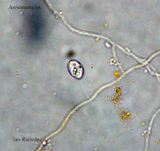
Picture 3. Anisonema sp. An anisonema can be seen with it's flagella somewhat hooked on the structure running diagnolly across the frame. It's second flagella can be seen as the dark spot in front of it. It was using the flagella in the front to pull or propel itself along while the longer flagella in the rear stablized it (Pennak, 1953, p. 47 Figure 21-B).


In general, the activity in my MicroAquarium this week seemed to be about the same as last week. I was able to observe relatively similar numbers in multicellular organisms like the euplotes and cyclops, while smaller unicellular organisms like the anisonema were slightly more abundant. Other than the anisonema, I was unable to identify any new organisms. 


Monday, October 28, 2013

Day 9 - October 25, 2013

My MicroAquarium hasn't had any drastic changes since Day 1. It seems the large unidentified nematode that was pictured and videotaped in my previous post has gone missing, and is presumed deceased. Additionally, there is a slight change in the color and perceived health of the moss and carnivorous plant. All three species of plants appear to be slightly less vibrant in color and struggling with the conditions of the system.

I was able to observe using both the microscope with a camera in place of the eyepiece lenses, and the microscope with the normal eyepiece lenses this week and both offered a unique perspective on the MicroAquarium. The microscope with normal dual eyepiece lenses allowed me to see into the system with a degree of depth that was truly amazing. I could see things like the fruit of the carnivorous plant Utricularia gibba L. in three dimensions and with enough depth and clarity to understand their shape and structure much better than with the camera based microscope used to take the pictures. I didn't see as many organisms using the microscope with normal eyepiece lenses because the magnification wasn't as strong, but I was able to see the three dimensional layout of the MicroAquarium and specifically, how the branches and leaves of the three plant species were integrated.

This week there were a few previously unseen organisms in my MicroAquarium:


Picture 1. Euplotes sp. An ovaled shaped euplotes cell with cilia on it's outer membrane and various inner structures (Patterson, 1996, p. 124 Figure 259).


Picture 2. Cyclops sp. A young cyclops lacking in egg sacks and with underdeveloped outer structures (Pennak, 1953, p. 384 Figure 241). 


Picture 3. Cyclops sp. A side-profile view of a young cyclops eating on the leaves of Amblestegium varium (Hedwig) Lindberg (McFarland, 2013)(Pennak, 1953, p. 384 Figure 241). 

In general, there seemed to be more activity in my MicroAquarium this week when compared to a week ago. While the large nematode was missing, there was no shortage of small single celled organisms, and even a multicellular Cyclops sp., to replace him. As far as I could tell,there were many Euplotes sp., but only one  Cyclops sp. visible in the whole system. 

In addition to the pictured organisms, I was able to observe and identify a number of Epalxis sp. in my MicroAquarium this week (Pennak, 1953, p. 141 Figure 311). The organism can best be described as asymetrical, but generally circular in shape, and as possessing many cilia on most of it's outer membrane. The cilia appeared to assist in it's movement and in moving debris towards it for processing. 

Bibliography

Bibliography

McFarland, Kenneth [Internet] Botany 111 Fall 2013. [cited October 28, 2013]. Available from http://botany1112013.blogspot.com/

Patterson DJ. 1996. Free-Living Freshwater Protozoa: A Colour Guide. Washington DC: ASM Press. 80p., 95p., 124p.,  and 173p.


Pennak RW. 1953. Fresh-Water Invertebrates of the United States. 1st ed. New York: The Ronald Press Company. 47 p., 141p., and 384 p.


Friday, October 18, 2013

Day 1 - October 16, 2013 - Microaquarium Setup!

Today we set up our aquariums and began to observe some of the activities taking place within them!


My aquarium was filled with water from the 7th site, Third-Creek in Tyson Park (Knoxville Co. Knoxville TN Partial shade exposure. N30 57 13.53 W83 56 32.37 824 ft. 10/14/2013) (McFarland 2013).

As seen in the above picture I filled the bottom of the aquarium with about 1/2cm of sediment, and then took the water from just above the sediment in the collected sample, and transferred it to make up around 2/3 of the water. Finally, I filled the last 1/3 of my aquarium with surface water from Dr. McFarland's collected sample from Third Creek in Tyson Park.

In addition to the water and sediment from Third Creek, my aquarium has two different types of mosses, and a carnivorous plant included in it's system.

The first moss is:

Amblestegium varium (Hedwig) Lindberg. Moss. Collection from: Natural spring. at Carters Mil Park, Carter Mill Road, Knox Co. TN. Partial shade exposure. N36 01.168 W83 42.832. 10/13/2013 (McFarland 2013).

The second moss is:

Fontinalis sp. Moss. Collected from: Holston River along John Sevier Hwy under I 40 Bridge Partial shade exposure Holston River water Shed N36 00.527 W83 49.549 823ft 10/13/2013 (McFarland 2013).

The carnivorous plant is:

Utricularia gibba L. Flowering plant. A carnivorous plant. Original material from south shore of Spain Lake (N 35o53 12.35" W088o20' 47.00), Camp Bella Air Rd. East of Sparta TN. in White Co. and grown in water tanks outside of greenhouse at Hesler Biology Building. The University of Tennessee. Knox Co. Knoxville, TN. 10/13/2013 (McFarland 2013).

The first moss, Amblestegium varium (Hedwig) Lindberg, can be identified as dark green color, while the second moss, Fontinalis sp., can be identified as a very dark reddish brown color. The carnivorous plant, Utricularia gibba L. is identified as being a light green with little black dots spread throughout the structure of the plant. These dots are dead flowers and will eventually be replaced with new growth. All three can be seen in the above picture of my aquarium (McFarland 2013).

Observations:

I was fortunate enough to observe my aquarium on the digital microscope during the very first observation session and got some really cool images and videos from it. Here are some of the pictures:


Picture 1. Unidentified Nematode moving violently.


Picture 2. Unidentified Nematode moving violently.


Picture 3. Unidentified organism moving violently.


Picture 4. Unidentified organism moving violently. 


I also got a few videos of activities:

and: